Can I Get Social Security Disability Benefits for Congestive (Chronic) Heart Failure?
- How Does the Social Security Administration Decide if I Qualify for Disability Benefits for Congestive Heart Failure?
- About Congestive Heart Failure and Disability
- Winning Social Security Disability Benefits for Congestive Heart Failure by Meeting a Listing
- Residual Functional Capacity Assessment for Congestive Heart Failure
- Getting Your Doctor’s Medical Opinion About What You Can Still Do
Winning Social Security Disability Benefits for Congestive Heart Failure by Meeting a Listing
To determine whether you are disabled at Step 3 of the Sequential Evaluation Process, the Social Security Administration will consider whether your congestive heart failure is severe enough to meet or equal the chronic heart failure listing. The Social Security Administration has developed rules called Listing of Impairments for most common impairments. The listing for a particular impairment describes a degree of severity that the Social Security Administration presumes would prevent a person from performing substantial work. If your chronic heart failure is severe enough to meet or equal the listing, you will be considered disabled.
The listing for chronic heart failure is listing 4.02, which has two parts, A and B. To meet the listing you must satisfy both part A and part B despite receiving prescribed treatment.
Part A is mainly concerned with diagnosis, while Part B deals with clinical and functional severity. A legitimate diagnosis of heart failure from any cause must be established before consideration of part A or part B of the listing. The critical question to determine listing-level severity is whether some degree of chronic heart failure remains after treatment for an acute episode of failure.
Meeting SSA Listing 4.02A for Heart Failure
You will meet Part A of Listing 4.02 if you have chronic heart failure (with characteristic symptoms and signs) while on a regimen of prescribed treatment and–
A. Medically documented presence of one of the following:
1. Systolic heart failure, with left ventricular end diastolic dimensions greater than 6.0 cm or ejection fraction of 30 percent or less during a period of stability (not during an episode of acute heart failure); or
2. Diastolic heart failure, with left ventricular posterior wall plus septal thickness totaling 2.5 cm or greater on imaging, with an enlarged left atrium greater than or equal to 4.5 cm, with normal or elevated ejection fraction during a period of stability (not during an episode of acute heart failure);
Part A.1: Systolic Heart Failure
Part A.1 requires systolic heart failure. Assuming that chronic heart failure is otherwise reasonably documented as a general requirement of the listing, part A.1 requires an objective determination of either cardiomegaly (cardiac enlargement) or left ventricular dysfunction.
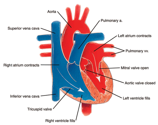
Cardiomegaly can be demonstrated with echocardiography showing a left ventricular end diastolic diameter of greater than 6.0 centimeters. This is actually the inside diameter of the left ventricular cavity during diastole—the left ventricular inside diastolic diameter (LVIDD). In diastole the heart is relaxed and filling with blood (see Figure 4 below).

Figure 4: The relaxed heart during diastole.
The LVIDD does not include the thickness of the heart muscle wall, because it is the dilation of the LV cavity that suggests a failing heart rather than the total diameter of the heart. Many hypertensive individuals have thickened heart muscle without heart failure (even though hypertension is associated with the risk of heart failure); in fact, even the thickness of a normal heart will be significantly greater than the LVIDD. Any other reliable cardiac imaging test, such as cardiac magnetic resonance imaging (cardiac MRI) or ventriculography performed during cardiac catheterization, can also satisfy the LVIDD measurement. However, echocardiography is the cheapest way to non-invasively get a good measurement of cardiac chamber size; contrast injection is not required for this type of measurement so no risk is involved.
It is also acceptable to document left ventricular dysfunction by showing left ventricular ejection fraction (LVEF) of 30% or less. The LVEF is the percent of blood in the left ventricle that the ventricle can pump out with each contraction. A normal LVEF is 55-65%. Most authorities would agree that an LVEF is not significantly abnormal until it falls below 50%. An LVEF of 30% or less provides the best quantified objective information and definitely shows serious heart disease. LVEF can be measured by any of the imaging studies mentioned.
LVEF alone does not always reveal chronic heart failure. A normal LVEF is usually not compatible with a diagnosis of systolic heart failure. In fact, an LVEF of more than 30% should make the diagnosis of chronic systolic heart failure suspect. But heart failure caused by diastolic dysfunction can be associated with a normal LVEF although there may be clinical signs of failure (in the present or past) in the form of venous congestion such as edema, hepatomegaly (enlarged liver), and jugular vein distention as well as shortness of breath with exertion and weakness. Fortunately, the Social Security Administration has added a provision for diastolic heart failure in Part A.2.
It is critical that the cardiac measurements be done during a period of stability after treatment for acute heart failure.
Part A.2: Diastolic Heart Failure
Part A.2 requires diastolic heart failure. The required abnormal cardiac measurements can most easily be obtained by echocardiography, but a cardiac MRI or any other reliable means of measuring cardiac dimensions are acceptable. The posterior muscular wall of the heart and interventricular partition separating the cardiac ventricles must be abnormally thickened to at least 2.5 centimeters (cm) and the left atrium must have a diameter of at least 4.5 cm. Since the LVEF in diastolic heart failure is normal or increased, that abnormality is also expected by part A.2. All of these measurements would be a routine part of any cardiac imaging study. In the medical literature, diastolic heart failure is also referred to as “heart failure with preserved ejection fraction.”
It is critical that the cardiac measurements be done during a period of stability after treatment for acute heart failure.
Meeting SSA Listing 4.02B for Heart Failure
You will meet Part B of Listing 4.02 if you have chronic heart failure (with characteristic symptoms and signs) while on a regimen of prescribed treatment–
B. Resulting in one of the following:
1. Persistent symptoms of heart failure which very seriously limit the ability to independently initiate, sustain, or complete activities of daily living in an individual for whom an MC, preferably one experienced in the care of patients with cardiovascular disease, has concluded that the performance of an exercise test would present a significant risk to the individual; or
2. Three or more separate episodes of acute congestive heart failure within a consecutive 12-month period, with evidence of fluid retention from clinical and imaging assessments at the time of the episodes, requiring acute extended physician intervention such as hospitalization or emergency room treatment for 12 hours or more, separated by periods of stabilization; or
3. Inability to perform on an exercise tolerance test at a workload equivalent to 5 METs or less due to:
a. Dyspnea, fatigue, palpitations, or chest discomfort; or
b. Three or more consecutive premature ventricular contractions (ventricular tachycardia), or increasing frequency of ventricular ectopy with at least 6 premature ventricular contractions per minute; or
c. Decrease of 10 mm Hg or more in systolic pressure below the baseline systolic blood pressure or the preceding systolic pressure measured during exercise due to left ventricular dysfunction, despite an increase in workload; or
d. Signs attributable to inadequate cerebral perfusion, such as ataxic gait or mental confusion.
If the Part A diagnostic considerations are satisfied, then any of B.1 through B.3 must also be fulfilled in regard to clinical/functional severity in order for you to meet the listing.
Part B.1 Persistent Symptoms
Part B.1 is satisfied by persistent symptoms consistent with chronic heart failure that are severe enough that a Social Security Administration medical consultant thinks cardiac exercise testing is contraindicated. Optimally, symptoms and limitations will be documented in your medical records and not mentioned simply in association with your application for disability benefits.
Part B. 2 Episodes of Acute Failure
Part B.2 requires at least 3 episodes of acute congestive heart failure (diastolic or systolic) in a consecutive 12-month period. By the requirement for “extended physician intervention such as hospitalization or emergency room treatment for 12 hours or more,” the Social Security Administration is assuring that most cases of repeated acute CHF will be easily identifiable.
The required evidence of fluid retention characteristic of acute congestive failure could be provided by hepatomegaly (liver enlargement), ascites (fluid in the abdomen), jugular vein distention or pressure, rales (abnormal lung sounds), peripheral edema (fluid retention and swelling in the extremities), or rapid weight gain.
The Social Security Administration correctly does not require that congestive signs be present on all examinations. In cases of prolonged ER treatment or hospitalization for acute CHF, it is usually easy to validate episodes of acute CHF based on history, physical examination, symptoms, and cardiac imaging. This kind of information is to be expected in the hospital record.
Part B.3 Inability to Perform on Exercise Tolerance Test
Part B.3 requires medical reasons related to chronic heart failure that caused you to be unable to achieve a workload equivalent to 5 METs or less (exertion approximately equal to brisk walking) during an exercise tolerance test.
Part B.3.a can be satisfied by inability to perform 5 METs exertion on an exercise test due to symptoms of heart failure, thus precluding the ability to do even sedentary work. Symptoms that indicate worsening heart failure developing during exercise are severe shortness of breath (SOB) and weakness, as well as chest discomfort such as angina pectoris and subjectively forceful heart beats (palpitations). Objective findings suggesting the onset of failure during exercise are the development of an S3 heart sound (an abnormal heart sound heard with a stethoscope that indicates ventricular dysfunction) or even frank pulmonary edema (fluid in the lungs). The pulmonary edema can be heard with a stethoscope as wet-sounding rales with inspiration, especially at the bottoms (bases) of the lungs.
In addition to symptoms of heart failure induced by exercise, part B.3.a can be satisfied by specific objective abnormalities that result in inability to do 5 METs of exertion. In most instances, these will be cases in which the treating physician has performed exercise testing.
Part B.3.b refers to abnormalities appearing on electrocardiogram (ECG, EKG) during exercise, in the form of abnormal beats. Part B.3.b describes an exercise-induced ventricular arrhythmia of a kind that can be life-threatening if it persists. Therefore, the exercise test must be stopped if these findings appear.
Part B.3.c refers to an abnormal blood pressure response to exercise, characterized as a fall of systolic pressure at least 10 mm Hg below the baseline, standing, pre-exercise level. A 10 mm Hg fall in systolic blood pressure at any time during exercise could also qualify. Such abnormality can be caused by a potentially life-threatening left ventricular dysfunction and requires immediate termination of exercise testing. However, in some situations a fall in blood pressure may be caused by more innocuous factors such as deconditioning, apprehension, and drugs. In these instances, as determined by informed medical judgment, part B.3.c would not be satisfied.
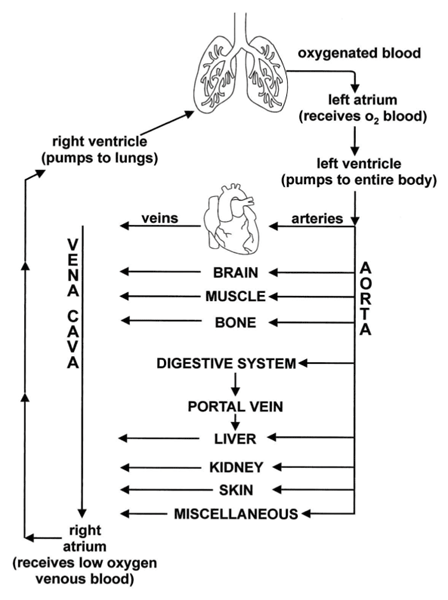
Part B.3.d recognizes the inability of a failing heart to supply the brain with blood during exertion (see Figure 5 below). This can result in the onset of an uncoordinated gait or mental confusion. The attending physician should immediately stop an exercise test in which these abnormalities appear, as they are life-threatening, or at least could cause a fall and serious injury.

Figure 5: Circulation of blood to the brain and other parts of the body.
Continue to Residual Functional Capacity Assessment for Congestive Heart Failure.
Go back to About Congestive Heart Failure and Disability.


