Can I Get Social Security Disability Benefits for Ischemic Heart Disease?
- How Does the Social Security Administration Decide if I Qualify for Disability Benefits for Ischemic Heart Disease?
- About Ischemic Heart Disease and Disability
- Winning Social Security Disability Benefits for Ischemic Heart Disease by Meeting a Listing
- Residual Functional Capacity Assessment for Ischemic Heart Disease
- Getting Your Doctor’s Medical Opinion About What You Can Still Do
Winning Social Security Disability Benefits for Ischemic Heart Disease by Meeting a Listing
To determine whether you are disabled at Step 3 of the Sequential Evaluation Process, the Social Security Administration will consider whether your ischemic heart disease is severe enough to meet or equal the ischemic heart disease listing. The Social Security Administration has developed rules called Listing of Impairments for most common impairments. The listing for a particular impairment describes a degree of severity that the Social Security Administration presumes would prevent a person from performing substantial work. If your ischemic heart disease is severe enough to meet or equal the listing, you will be considered disabled.
The listing for ischemic heart disease is listing 4.04, which has three parts, A, B, and C. To meet the listing you must satisfy any one of the three parts despite receiving prescribed treatment.
Symptoms Due to Myocardial Ischemia
To satisfy any of the three parts of the ischemic heart disease listing, you must have symptoms of myocardial ischemia. These are:
- Typical angina pectoris. This is chest pain brought on by effort or emotion and promptly relieved by rest, sublingual nitroglycerin (that is, nitroglycerin tablets that are placed under the tongue), or other rapidly acting nitrates. Typically, the discomfort is located in the chest (usually under the breast bone) and described as pressing, crushing, squeezing, burning, aching, or oppressive.
- Atypical angina. This is discomfort or pain from myocardial ischemia that is felt in places other than the chest. The common sites of cardiac pain are the inner left arm, neck, jaw, upper abdomen, and back, but the discomfort or pain can be elsewhere. To represent atypical angina, your discomfort or pain should have precipitating and relieving factors similar to those of typical chest discomfort.
- Anginal equivalent. This means shortness of breath (dyspnea) on exertion without chest pain or discomfort. Your shortness of breath should have precipitating and relieving factors similar to those of typical chest discomfort. In these situations, it is essential to establish objective evidence of myocardial ischemia to ensure that you do not have effort dyspnea due to non-ischemic or non-cardiac causes.
- Variant angina. Variant angina (Prinzmetal’s angina, vasospastic angina) refers to the occurrence of anginal episodes at rest, especially at night, accompanied by transitory ST segment elevation (or, at times, ST depression) on an ECG. It is due to severe spasm of a coronary artery, causing ischemia of the heart wall, and is often accompanied by major ventricular arrhythmias, such as ventricular tachycardia (rapid heart beat).
- Silent ischemia. Myocardial ischemia, and even myocardial infarction, can occur without pain or any other symptoms. Pain sensitivity may be altered by a variety of diseases, most notably diabetes mellitus and other neuropathic disorders. Individuals also vary in their threshold for pain.
See Angina Pectoris and Ischemic Heart Disease.
Although the whole listing has a basic requirement for angina, a finding that your impairment equals the listing might be appropriate, if the other combined symptoms are just as limiting in the absence of any angina.
Meeting Social Security Administration Listing 4.04A for Ischemic Heart Disease
You will meet Part A of Listing 4.04A if you have ischemic heart disease, with symptoms due to myocardial ischemia, while on a regimen of prescribed treatment, with:
A. Sign-or-symptom limited exercise test demonstrating at least one of the following manifestations at a workload equivalent to 5 METs or less:
1. Horizontal or down-sloping depression, in the absence of digitalis glycoside treatment or hypokalemia, of the ST segment of at least -0.10 millivolts (-1.0 mm) in at least 3 consecutive complexes that are on a level baseline in any lead other than aVR, and depression of at least -0.10 millivolts lasting for at least 1 minute of recovery; or
2. At least 0.1 millivolt (1 mm) ST elevation above resting baseline in non-infarct leads during both exercise and 1 or more minutes of recovery; or
3. Decrease of 10 mm Hg or more in systolic pressure below the baseline blood pressure or the preceding systolic pressure measured during exercise (see §4.00E9e) due to left ventricular dysfunction, despite an increase in workload; or
4. Documented ischemia at an exercise level equivalent to 5 METs or less on appropriate medically acceptable imaging, such as radionuclide perfusion scans or stress echocardiography.
Part A concerns abnormalities appearing during exercise stress testing. Exercise testing is important in disability determination, because exercise can unmask cardiac ischemia that is not present at rest.
METS
The abnormalities listed in Part A must appear at a level of exercise (workload) that is equivalent to an estimated 5 METs (metabolic equivalents ) or less. Metabolic equivalents are a measure of oxygen use per unit body weight per minute. One MET is the body’s use of 3.5 milliliters of oxygen for each kilogram4 of body weight per minute. METs are the standard for expressing how much exertion a person is capable of performing. The higher the physical workload, the higher the MET level required, because more oxygen must used. Activities requiring approximately 5 METs include brisk walking, raking leaves, and light carpentry.
Exercise Test Results
These test results could be obtained from your medical records or through a consultative examination that the Social Security Administration arranges for you. The Social Security Administration should send you for stress testing only when necessary to determine disability. Non-medical doctors, such as disability examiners, claim managers, or disability hearing officers, are unqualified to determine when stress testing is indicated or to evaluate the results.
The Social Security Administration will not use interpretations of resting or exercise ECGs without the actual tracings, or legible copies, for review. Furthermore, the complete report of the test should be available, including the protocol used (time per stage, as well as speed and tilt of a treadmill, for example), blood pressure measurements during testing, and description of any symptoms or physical abnormalities noted during testing. Unfortunately, many cardiac exercise tests are performed in hospitals and hospitals tend to throw away the tracings, only keeping the written narrative of the test results. If so, it is impossible for the Social Security Administration to validate the ECG findings.
Angina
Although angina is a general requirement of the listing, angina does not have to be induced during exercise testing for part A to be satisfied. However, angina may occur during testing and be relieved promptly by cessation of exercise and administration of sublingual nitroglycerin under the observation of a physician. This event would further strengthen the diagnosis of very limiting ischemic heart disease.
Parts A.1 and A.2 Technical Electrical Abnormalities
Parts A.1 and A.2 specify the technical electrical abnormalities that must be present on your ECG at 5 METs or less to meet the listing. The hallmark of ischemia on ECG is what is called ST segment depression, a phrase that is frequently seen in the medical records of individuals with ischemic heart disease. Ischemia can also be demonstrated by an elevation of the ST segment (part A.2), although this is less common.
An ECG is a line that changes in accordance with one cycle of the heart’s electrical activation and recovery, i.e., one heart beat. An actual ECG plots a line of these cycles, one after the other. The standard ECG has 12 leads, which means that 12 different electrodes are placed over different parts of the central and left chest and each records a slightly different view of the heart’s electrical activity.
A resting ECG is the least sensitive method of detecting cardiac ischemia. An ECG done during exercise is much more likely to provoke ischemia, but is still much less effective than testing done with both ECGs and some type of imaging study of the heart during exercise, like an exercise radionuclide study (e.g., thallium treadmill stress test) or exercise echocardiogram (part A.4).
Part A.3 Inability to Increase Systolic Blood Pressure
Part A.3 recognizes that an inability to adequately increase systolic blood pressure (SBP) with exercise even at a low 5 MET level implies severe heart disease, because the heart cannot pump well enough to maintain blood pressure.
This is the same finding required by listing 4.02B3c.
Part A.4 Radionuclide Exercise Testing
Thallium-201 is an example of a radioactive isotope that can be used with exercise testing; technetium is another frequently used radionuclide. Thallium emits x-rays and is not taken up as well by heart tissue that has a poor blood flow (perfusion defect), relative to normal parts of the heart. When the chest is scanned, an image can be constructed from the x-rays. Ischemic perfusion defects are reversible—they appear with exertion and disappear at rest. Fixed defects may indicate scar tissue form a prior heart attack, but can also be “stunned” muscle that is not actually irreversibly dead.
Combining thallium with exercise ECGs—whether a treadmill or bicycle ergometer—greatly increases accuracy in detecting ischemia. The Social Security Administration does not require the actual test films showing a perfusion; the full report and medical interpretation are sufficient. The Social Security Administration refers to “documented” thallium abnormalities, and means an expert interpretation by a cardiologist or radiologist. Second-hand reports of test results written in treating source medical records are not reliable. If judged necessary for accurate evaluation, Social Security Administration medical consultants can request that you undergo exercise testing with radionuclides such as thallium.
If a stress radionuclide study is done, there will always be accompanying ECGs. This situation leaves several possibilities:
1. Both the radionuclide perfusion study and the ECGs are negative for ischemia.
2. Both the perfusion study and the ECGs are positive for ischemia.
3. The perfusion study is positive for ischemia, but the ECGs are negative for ischemia.
4. The perfusion study is negative for ischemia, but the ECGs are positive for ischemia.
5. The perfusion study and/or ECGs are equivocal for ischemia.
Possibilities 1 and 2 could reliably be interpreted as normal and abnormal studies, respectively. Possibility 3 should be interpreted as an abnormal study, because one of the reasons a perfusion study is done along with ECG tracings is that an ECG alone may not be sensitive enough to detect significant ischemia.
Possibility 4 is a little more problematic. Although it is possible to have false negative perfusion results in rare instances, it is much more likely that the ECG tracings are false positive. This is particularly true in electrolyte imbalances, presence of drugs such as digitalis, or women (natural or supplemental estrogens can cause false positive ECG changes).
Possibility 5 requires the most medical judgment; other evidence of heart disease should have considerable weight in these instances. The ability to make judgments regarding test validity is implicit in disability adjudication. Obviously, this judgment should be done only by individuals with professional medical training.
Resting ECG Abnormalities
Part A does not mention the possibility of an abnormal resting ECG showing cardiac ischemia. A resting ECG performed while a person has angina will show myocardial ischemia about half the time.
If you have chest pain compatible with angina and resting ECG changes that are compatible with ischemia, the Social Security Administration should give serious consideration to a finding of equivalent severity to part A without requiring exercise testing. A concurrent exercise test already in the medical evidence that shows no ischemia would take priority over resting changes, because true ischemia on a resting ECG would worsen with exertion.
Medical judgment must be applied on a case by case basis in regard to how resting ECG abnormalities are adjudicated.
Meeting Social Security Administration Listing 4.04B for Ischemic Heart Disease
You will meet Part B of Listing 4.04 if you have ischemic heart disease, with symptoms due to myocardial ischemia, while on a regimen of prescribed treatment, with three separate ischemic episodes, each requiring revascularization or not amenable to revascularization, within a consecutive 12-month period.
Part B recognizes that repeated episodes of clinical deterioration as a result of ischemic heart disease can be disabling.
To count as an episode you must have had revascularization either by angioplasty (with or without stenting) or bypass surgery. If surgery is not done, the ischemic episode can still qualify if not amenable to surgery. “Not amenable” means that the revascularization procedure could not be done because of another medical impairment or because the vessel was not suitable for revascularization. Re-occlusion of a vessel during the same hospitalization does not count for listing purposes.
If you satisfy this part of the listing, you could be found to have had significant medical improvement and no longer be disabled at a future continuing disability review once your condition has stabilized, if you don’t meet any other part of this listing or another listing.
Meeting Social Security Administration Listing 4.04C for Ischemic Heart Disease
You will meet Part C of Listing 4.04 if you have ischemic heart disease, with symptoms due to myocardial ischemia, while on a regimen of prescribed treatment, with:
- Coronary artery disease,
- Demonstrated by angiography (obtained independent of Social Security disability evaluation) or other appropriate medically acceptable imaging, and
- In the absence of a timely exercise tolerance test or a timely normal drug-induced stress test,
- An MC, preferably one experienced in the care of patients with cardiovascular disease, has concluded that performance of exercise tolerance testing would present a significant risk,
- With both 1 and 2:
1. Angiographic evidence revealing:
a. 50 percent or more narrowing of a non-bypassed left main coronary artery; or
b. 70 percent or more narrowing of another non-bypassed coronary artery; or
c. 50 percent or more narrowing involving a long (greater than 1 cm) segment of a non-bypassed coronary artery; or
d. 50 percent or more narrowing of at least 2 non-bypassed coronary arteries; or
e. 70 percent or more narrowing of a bypass graft vessel; and
2. Resulting in very serious limitations in the ability to independently initiate, sustain, or complete activities of daily living.
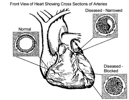
Coronary artery disease (CAD) is by far the most common cause of ischemic heart disease (see Figure 6 below). The term is understood to refer to obstructive lesions in the largest coronary arteries, known as the epicardial arteries because they lie near the surface of the heart. These arteries arise from the aorta just as blood is ejected from the left ventricle through the aortic valve into the systemic circulation, so that the heart itself is the first organ to receive oxygenated blood.

Figure 6: Detail of arteries in a normal heart and in a heart with coronary artery disease.
Part C applies only when no timely exercise tolerance test (ETT) or other cardiac stress test is available and when a Social Security medical consultant concludes that stress testing would present a significant risk to the claimant. If these conditions are fulfilled, then Part C has both angiographic criteria (C.1a-e) and functional loss criteria (C.2). Both must be satisfied.
Part C.1 Angiographic Criteria
A re-obstructed artery following prior angioplasty or bypassing is non-bypassed for purposes of the listing.
In prior listings, the Social Security Administration would have required cardiac catheterization done independently of the Social Security Administration to demonstrate the severity of coronary artery lesions. Currently, less invasive procedures are fully satisfactory imaging modalities, including electron beam computed tomography, high-resolution CT scanning, and magnetic resonance imaging. In some instances, echocardiography can be used to image parts of larger coronary arteries. Of course, the Social Security Administration does not have to accept inappropriate imaging that does not reveal the nature, location, and severity of the lesions sufficiently to allow reasonable application of the listing.
The new high-resolution 64-slice CT scanners are capable of providing excellent imaging of coronary arteries and are increasingly being used to screen patients who have chest pain for coronary artery disease. Although some radiation is delivered to the patient, such CT angiography can provide a faster and more cost-effective way for diagnosing coronary disease as the probable cause of chest pain, compared to stress testing with radionuclides or conventional angiography by cardiac catheterization. As the resolution of cardiac MRI scanners improves, it is likely that the future will permit imaging of the heart without any X-rays. Conventional angiography exposes the patient to X-rays, as well as being invasive and more risky and expensive. However, if intervention is eventually needed (such as stenting a coronary artery), then cardiac catheterization is unavoidable.
Part C.2 Functional Loss Criteria
Part C.2 provides guidance on functional loss as a result of limitations caused by ischemic heart disease. At this level of severity, a claimant would normally have significant amounts of medical records, including hospitalization records to support the credibility of markedly limiting symptoms: fatigue, palpitation, dyspnea (shortness of breath), or angina. Angina alone is a sufficient symptom for allowance under part C.2, if severe enough. If not, additional symptoms of fatigue, palpitations, or dyspnea combined with angina, are sufficient for allowance under part C.2.
It cannot be over-emphasized that specific examples of limitations in activities of daily living are much more helpful to the Social Security Administration than generalizations.
Continue to Residual Functional Capacity Assessment for Ischemic Heart Disease.
Go back to About Ischemic Heart Disease and Disability.


